Routine Non- Invasive Diagnostic Testing & Therapeutic Cardiology Out Patient Procedures

Ankle-brachial index test

ANKLE-BRACHIAL INDEX TEST

Ankle-brachial index test is a comparison of your blood pressure measured at an ankle and your blood pressure measured at an arm, which can indicate blockage of blood flow to the legs.

After removing your socks and shoes you will lie on an examining bed. The technician will attach blood pressure cuffs to arms, ankles, and toes. The sonographer will warn you when the cuffs inflate. 

You can take the following steps to prepare for your appointment:

  • DO NOT wear a long sleeve shirt.
  • DO NOT wear pantyhose
  • An ankle brachial index usually takes about 10 to 15 minutes.
Echocardiogram

ECHOCARDIOGRAM

An echocardiogram uses sound waves to produce images of your heart. This commonly used test allows your doctor to see how your heart is beating and pumping blood. Your doctor will look for heart size, pumping strength, damage to the heart muscle, valve problems, and heart defects.

Transthoracic echocardiogram is a standard, noninvasive echocardiogram. A technician (sonographer) spreads gel on your chest and then presses a device known as a transducer firmly against your skin, aiming an ultrasound beam through your chest to your heart. The transducer records the sound wave echoes your heart produces. A computer converts the echoes into moving images on a monitor. Doppler echocardiogram is when sound waves bounce off blood cells moving through your heart and blood vessels, they change pitch. These changes (Doppler signals) can help your doctor measure the speed and direction of the blood flow in your heart. Doppler techniques are used in most transthoracic echocardiograms, and can check blood flow problems and blood pressures in the arteries. 

After undressing from the waist up, you'll lie on an examining bed. The technician will attach sticky patches (electrodes) to your body to help detect and conduct the electrical currents of your heart. During the echocardiogram, the technician will dim the lights to better view the image on the monitor. You may hear a pulsing "whoosh" sound, which the machine is recording the blood flowing through your heart. Most echocardiograms take 45 minutes, but the timing may vary depending on your condition. During a transthoracic echocardiogram, you may be asked to breathe in a certain way or to roll onto your left side. Sometimes the transducer must be held very firmly against your chest. This can be uncomfortable — but it helps the technician produce the best images of your heart.

No special preparations are necessary for a standard transthoracic echocardiogram.

Carotid Ultrasound

CAROTID ULTRASOUND

The primary purpose of a carotid ultrasound is to screen for a narrowing of the carotid arteries that indicates an increased risk of stroke. Narrowing is usually caused by plaques — a buildup of fats, cholesterol, calcium and other substances that circulate in the bloodstream. Early detection of narrowing of the carotid arteries enables your doctor to begin treatments that improve blood flow to the brain and decrease your risk of stroke. 

Other uses of carotid ultrasound: your doctor also may order a carotid ultrasound to:

  1. Evaluate the structure and function of the artery after surgery to remove plaques (carotid endarterectomy)
  2. Evaluate the placement and treatment effect of a stent, a mesh tube used to improve blood flow through an artery and support a weakened artery
  3. Detect other abnormalities in the structure of a carotid artery that may disrupt blood flow

You can take the following steps to prepare for your appointment:

  1. Wear a comfortable shirt with no turtleneck and don’t wear a necklace.

During the procedure you'll likely lie down on your back during the procedure. The ultrasound technician (sonographer) may gently adjust the position of your head to improve access to the side of your neck. The sonographer will apply a warm gel to your skin above the site of each carotid artery. The gel helps eliminate the formation of air pockets between your skin and the transducer. The sonographer then gently presses the transducer against the side of your neck in order for the instrument to send and receive sound waves. You shouldn't feel any discomfort during the procedure. If you do, tell the sonographer. A carotid ultrasound usually takes about 15 to 30 minutes.

Vascular ultrasound

VASCULAR ULTRASOUND (ARTERIAL & VENOUS ULTRASOUND)

Vascular ultrasound is a noninvasive ultrasound method (also called a duplex study) used to examine the blood circulation in the arms and legs. Non- invasive means the procedure does not require the use of needles, dyes, radiation or anesthesia.

During a vascular ultrasound, sound waves are transmitted through the tissues of the area being examined. These sound waves reflect off blood cells moving within the blood vessels, allowing the reading physician to calculate their speed. The sound waves are recorded and displayed on a computer screen.

Your physician has recommended that you have this test to evaluate the blood flow to specific part in your body. Vascular ultrasound can be used to evaluate:

  • Locate and identify blockages (stenosis) and abnormalities like plaque or emboli and help plan for their effective treatment. 
  • Evaluate the success of procedures that graft or bypass blood vessels.
  • Determine if there is an enlarged artery (aneurysm).
  • Detect blood clots (deep venous thrombosis (DVT) in the legs or arms.
  • Determine the source and severity of varicose veins.

Your ultrasound test is performed by specially trained technologists and interpreted by your cardiologist. You will lie on the bed during the test. A small amount of water-soluble gel is applied to the skin over the area to be examined. The gel does not harm your skin or stain your clothes. A small device called a transducer is placed over the gel-coated area to be examined. The transducer produces images on the ultrasound screen. There may be some minor discomfort during the exam when the technologist applies pressure to your arms or legs. You may hear noises when the technologist listens to the blood flow and records the measurements. 

The ultrasound takes about 30 to 45 minutes to complete. 

There is no special preparation before the test

Stress echocardiogram

STRESS ECHOCARDIOGRAM

Some heart problems — particularly those involving the coronary arteries that supply blood to your heart muscle — occur only during physical activity. For a stress echocardiogram, ultrasound images of your heart are taken before and immediately after walking on a treadmill. If you're unable to exercise, you may get an injection of a medication to make your heart work as hard as if you were exercising.

After undressing from the waist up and putting a gown on. The treadmill technician will attach sticky patches (electrodes) to your body to help detect and conduct the electrical currents of your heart. The sonographer will obtain images of your heart (left ventricle) before getting on the treadmill. The cardiologist will then be present when you're on the treadmill. We will encourage you to do your best, if you have trouble on the treadmill just tell us. We are trying to get your heart rate to a certain point. Once you reach that point we ask that you lie on the bed and the sonographer will obtain images of your heart beating fast.

During a stress echocardiogram, exercise or medication — not the echocardiogram itself — may temporarily cause an irregular heartbeat. Serious complications, such as a heart attack, are rare.

If you'll be walking on a treadmill during a stress echocardiogram, you should wear:

  • Comfortable clothing
  • Don’t wear a dress
  • Comfortable shoes
  • No heels 

DUPLEX ULTRASOUND

A duplex ultrasound is a test to see how blood moves through your arteries and veins.

How the Test is Performed

A duplex ultrasound combines:

  • Traditional ultrasound: This uses sound waves that bounce off blood vessels to create pictures.
  • Doppler ultrasound: This records sound waves reflecting off moving objects, such as blood, to measure their speed and other aspects of how they flow.

There are different types of duplex ultrasound exams. Some include:

  • Arterial and venous duplex ultrasound of the abdomen. This test examines blood vessels and blood flow in the abdominal area.
  • Carotid duplex ultrasound looks at the carotid artery in the neck.
  • Duplex ultrasound of the extremities looks at the arms or legs.
  • Renal duplex ultrasound examines the kidneys and their blood vessels.

You may need to wear a medical gown. You will lie down on a table, and the ultrasound technician will spread a gel over the area being tested. The gel helps the sound waves get into your tissues.

A wand, called a transducer, is moved over the area being tested. This wand sends out the sound waves. A computer measures how the sound waves reflect back, and changes the sound waves into pictures. The Doppler creates a "swishing" sound, which is the sound of your blood moving through the arteries and veins.

You need to stay still during the exam. You may be asked to lie in different body positions, or to take a deep breath and hold it.

Sometimes during a duplex ultrasound of the legs, the health care provider may calculate an ankle-brachial (ABI) index. You will need to wear blood pressure cuffs on your arms and legs for this test.

The ABI number is obtained by dividing the blood pressure in the ankle by the blood pressure in the arm. A value of 0.9 or greater is normal.

How to Prepare for the Test

Usually, there is no preparation for this test.

If you are having an ultrasound of your stomach area, you may be asked not to eat or drink after midnight. Tell the person doing the ultrasound exam if you are taking any medicines, such as blood thinners. These might affect the results of the test.

How the Test will Feel

You may feel some pressure as the wand is moved over the body, but there is no discomfort most of the time.

Why the Test is Performed

A duplex ultrasound can show how blood flows to many parts of the body. It can also tell the width of a blood vessel and reveal any blockages. This test is a less invasive option than arteriography and venography.

A duplex ultrasound can help diagnose the following conditions:

  • Abdominal aneurysm
  • Arterial occlusion
  • Blood clot
  • Carotid occlusive disease (See: Carotid duplex)
  • Renal vascular disease
  • Varicose veins
  • Venous insufficiency

A renal duplex ultrasound can also be used after transplant surgery. This shows how well a new kidney is working.

ultrasound of the aorta

ULTRASOUND OF THE AORTA

Your doctor may recommend you have an ultrasound of the aorta if you're at risk of an abdominal aortic aneurysm. Aortic ultrasound screens for a potential abdominal aortic aneurysm, an enlargement or ballooning of the large artery that supplies blood to the abdomen, pelvis and legs.

Risk factors for abdominal aortic aneurysm include:

  • Being Male
  • Age of 60 years or older
  • A history of atherosclerosis-a thickening of the walls of your arteries
  • A family history of abdominal aortic aneurysm
  • Smoking
  • High blood pressure (a systolic pressure of 140 to 159 millimeters of mercury or mm Hg, and a diastolic pressure of 90 to 99 mm Hg
  • An aneurysm in one of your other arteries
  • Chronic lung disease

Food and liquids in your stomach can make it difficult for the ultrasound technician (sonographer) to get a clear picture of your aorta. Fasting instructions will be given before the exam. 

DAY BEFORE EXAMINATION: Clear liquids at noon and evening meals. Eat no solid foods. Drink four 8-ounce glasses of water between 1:00 p.m. and 9:00 p.m. 

At 5:00 p.m., drink one bottle (10 ounces) of Citrate of Magnesia. 

At 8:00 p.m., take two Biscodyl (Dulcolax) pills. 

Nothing to eat or drink after midnight (may take medications with water at anytime).

Nuclear stress test

NUCLEAR EXCERCISE STRESS TEST

What is a nuclear exercise stress test?

A nuclear exercise stress test is a diagnostic test used to evaluate blood flow to the heart. During the test, a small amount of radioactive tracer is injected into a vein. A special camera, called a gamma camera, detects the radiation released by the tracer to produce computer images of the heart.
Combined with exercise, the test can help determine if there is adequate blood flow to the heart during activity versus at rest. 

Can I eat or drink on the day of the test?

Yes. However, DO NOT eat or drink anything but water for 4 hours before the test. If you must take medications, drink only drink water to help you swallow your pills.
Avoid all products containing caffeine for 24 hours before the test. In general, caffeine is found in coffee, tea, colas, Mountain Dew®,chocolate and decaffeinated products. Decaffeinated products contain trace amounts of caffeine.

Should I take my medications the day of the test?

Please bring all of your medications, including over-the-counter medications and supplements that you routinely take, to the test appointment.
Please follow these guidelines about taking your medications the day of the test.
  • Medications with caffeine: DO NOT take any over-the-counter medication that contains caffeine (such as Excedrin®, Anacin®, diet pills and No Doz®) for 24 hours before the test. Ask your physician, pharmacist or nurse if you have questions about other medications that may contain caffeine. 
  • If you have asthma: Please bring your asthma inhaler medication to the test. 
  • If you have diabetes: If you take medications to control your blood sugar, do not take your medication until after the test is complete. Bring your diabetes medications with you so you can take it when the test is complete. Do not take your diabetes medication and skip a meal before the test. Plan to eat and take your blood sugar medication following your test. 

What should I wear for the test?

  • Please wear comfortable 2 piece clothing and shoes suitable for walking during the test. Ladies, it's preferable you not wear dresses or skirts.  
  • Please DO NOT wear sandals.
  • Do not wear any clothing that has metal on the shirt. This includes metal snaps on shirts.
Please DO NOT bring children with you to the testing. We are using radiation for your testing and there will be several other patients like yourself here. Therefore, you will not want children or anyone pregnant exposed unnecessarily to the radiation.

What to expect during the test

The testing area is supervised by a physician. 
A nuclear medicine technologist will place an IV into a vein in your arm or hand and inject a small amount of radioactive tracer. The tracer is not a dye or contrast. After the tracer is injected, you will wait approximately 1 hour before the first set of “resting” images are taken. 
Then you will be asked to lie very still under the gamma camera with both arms above your head for about 7 minutes. The camera will record images that show blood flow through your heart at rest. 
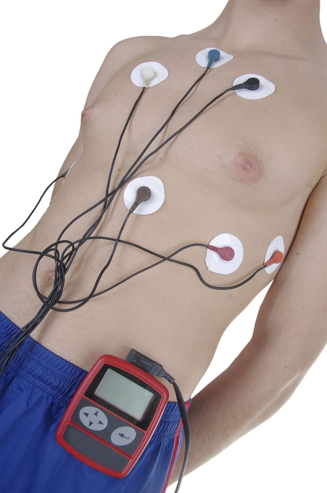
Next, a technician will place electrodes on your chest to monitor your EKG while you exercise. 
You will walk on a treadmill. At regular intervals, the difficulty of the exercise will increase until you achieve the target heart rate. Then, a second dose of radioactive tracer will be injected into the IV. Your heart rate, EKG and blood pressure will be monitored throughout the test. If you are unable to achieve your target heart rate or walk on a treadmill, a medication will be given to simulate exercise. 
After you have exercised, you will be offered a snack of coffee or juice and crackers. You may also bring a snack from home, if you prefer. Caffeine is permitted AFTER you have exercised.
About 45 minutes after exercising, you will be asked to again lie very still under the camera with both arms over your head for about 5 minutes. The camera will record images that show blood flow through your heart during exercise. These images will be compared to the first set. 

How long will the test last?

The appointment will take about 3 to 4 hours. The actual exercise part of the test usually lasts about 5-12 minutes.

What if I need to reschedule my test?

If you are unable to keep your appointment, please notify our office at least 24 hours in advance. Failure to do so may result in forfeiture of your deposit.

How do I get the results of my test?

After the cardiologist reviews your test, the results will go into your electronic medical record. Your referring physician will have access to the results and will contact you to discuss them.


HOLTER MONITOR

What is a Holter monitor?

A Holter monitor is a battery-operated portable device that measures and tape records your heart’s activity (ECG) continuously for 24 to 48 hours or longer depending on the type of monitoring used. The device is the size of a small camera. It has wires with silver dollar-sized electrodes that attach to your skin. The Holter monitor and other devices that record your ECG as you go about your daily activities are called ambulatory electrocardiograms.

Quick facts:

  • You may be asked to wear a Holter monitor to see if you have a slow, fast or irregular (uneven) heartbeat. Or, your doctor may use it to see how well your medicines are working to treat these problems. If you have a pacemaker and feel dizzy, your doctor may use a Holter monitor to find out if your pacemaker is working properly.
  • This monitor has no risks and wearing it isn’t painful.
  • The results of wearing a Holter monitor will help you and your doctor decide if you need more tests or medicines for your heart, or if you need a pacemaker or cardioversion procedure to restore a regular heart rhythm.

“I thought wearing the monitor at night would be very uncomfortable, but the cord was long so I could put it on the nightstand. I made sure the electrodes were on tight before I went to bed.” Sarah, age 62.

Why do people wear Holter monitors?

Regular electrocardiograms (ECGs or EKGs) let your doctor look at your heart’s activity at one point in time during your ECG test. But abnormal heart rhythms and cardiac symptoms may come and go. That’s why your doctor may want to evaluate your heartbeat over time while you go about your normal activities. You may be asked to wear a Holter monitor if you have fast, slow or irregular heartbeats called arrhythmias.

Wearing the monitor may tell your doctor:

  • If your medicines are working.
  • Why you have symptoms such as dizziness, faintness or the feeling that your heart is racing or skipping a beat.
  • If your heart is getting enough oxygen to meet its needs.

What are the risks of Holter monitors?

Wearing a Holter monitor has no risks and causes no pain. Holter monitors have wires that connect to small discs (electrodes) attached to your chest to record the electrocardiogram. Because the electrodes are attached with tape or adhesives, they may cause mild skin irritation. Tell the technician if you are allergic to any tapes or adhesives.

What should I expect with the Holter monitor?

A specially trained technician will attach the Holter monitor and instruct you how to record your symptoms while wearing it.

  • The technician first attaches the electrodes to your chest. If you have a hairy chest, he or she may shave some hair off to attach the electrodes firmly.
  • Once the electrodes are in place, the technician helps you put the Holter monitor on and explains how to take care of it. 
  • You can carry the monitor in a pocket or pouch, slung across your shoulders and neck like a purse or camera, or attach it to your waist.

Do your usual activities while you wear the monitor with these exceptions:

  • Don’t bathe, shower or swim while wearing the monitor.
  • Don’t have X-rays while wearing the monitor.
  • Stay away from high-voltage areas, metal detectors or large magnets.
  • The technician will show you how to keep a diary of your activities and symptoms during the test. It’s important to keep an accurate diary. If you feel symptoms such as chest pain, shortness of breath, uneven heartbeats or dizziness, note in your diary the time of day they began and what you were doing. Your diary will be compared to the changes in your ECG recorded by the Holter monitor.

What happens after wearing a Holter monitor?

After the test period, return the monitor to the technician. He or she will process the record of your heart activity and prepare a report for your doctor. This will include the notes in your diary. You should get the results of the test in one or two weeks.

ADDITIONAL SERVICES

We provide the following:

  • 24 Hour Cardiac Services
  • Preventive Cardiology
  • Cardiovascular Disease Management
  • Cardiac Catheterization
  • Coronary Angioplasty
  • Cardiac Computer Tomography
  • Stress Testing
  • 24 Hour Holter Monitoring
  • Echocardiography, Doppler Echocardiography
  • Peripheral Vascular Diagnostic Testing
  • Doppler Echocardiography
  • Transesophageal Echocardiography
  • Nuclear Cardiology
  • Arrhythmia Management
  • Peripheral Artery Disease (PAD) Management
  • Carotid Stent Grafting
  • Aortic Endovascular Stent Grafting
  • Temporary and Permanent Pacemakers
  • Electrophysiology
  • Pacemaker Clinic
  • Lipid Clinic
  • Coumadin Clinic
  • Congestive Heart Failure Clinic

Patient Information - FAQs

If you have questions about common cardiac terms, we highly recommend the American Heart Association A-Z guide for heart disease and stroke victims. This glossary will provide a useful reference for patients and their families. In addition, this page also provides important bio-statistical information for the medically curious.

American Heart Association A-Z Guide

American Heart Association A-Z Guide

AHA Scientific Statements and Guidelines

AHA Scientific Statements and Guidelines

American Heart Association website

American Heart Association website

The American Heart Association's Nutrition Center

The American Heart Association's Nutrition Center

The American Heart Association's Guide to Stress Management

The American Heart Association's Guide to Stress Management 

American Diabetes Association

American Diabetes Association

EMERGENCIES

Emergency calls can be avoided with foresight. Treatment of a condition before it becomes an emergency is the surest way to better health and few hospitalizations. 

It is always best to call your regular physician before going to the Emergency Room, since many hospital visits can be avoided. If you are unsure of whether you have an emergency please call your regular physician to help you determine this. If you are unable to reach your regular physician during a cardiac emergency, you may reach us or our covering physician by calling the office. If your situation does not allow time for a return phone call from the physician please go to the nearest hospital Emergency Room where a 24 hour-a-day Emergency Room Physician can evaluate you quickly and determine the stability of your situation.

FEES AND INSURANCE

Fees for medical services at our office will be due and payable at the time of your visits. By asking you to do this we can cut down the cost of billing and bookkeeping; and hopefully, keep your medical fees down. Our office, of course, will assist you when requested in helping you to collect your entitled reimbursement to the best of their ability. 

Our billing office will file all claims to third party contractors after you meet your co-pay and deductible. Should you have any questions concerning your account, please call our billing office number at (361) 887-2970.

OFFICE HOURS AND APPOINTMENTS

We see patients in the office who have either been referred by a personal physician or who have been seen previously in the hospital. Our office staff is available from 9:00 a.m. to 5:00 p.m. Monday thru Friday. We see patients by appointment only from 9:00 a.m. to 4:00 p.m. Monday thru Friday.


We ask that all HMO patients be responsible for obtaining their Primary Care Physician (PCP) referral forms prior to scheduling an appointment. We make every effort to honor all time commitments and request that you extend the same courtesy to us. On occasion, emergencies can cause problems, and whenever possible, you will be fully informed if there will be any delays because of this.

FOLLOW US

SHARE THIS PAGE!

Coastal Cardiology Logo

COASTAL CARDIOLOGY PLLC LOCATIONS

MAIN OFFICE

613 Elizabeth Street, Suite 402
Corpus Christi, Texas 78404
(361) 887-2900
Fax (361)887-0942

REFERRALS
Please fax new patient referrals to (361)900-3531 or email them to referrals@ccardiology.com

Diagnostic Testing fax number (361)887-2748

Riverside office fax number (361)933-0717

CALALLEN OFFICE
13725 Northwest Blvd., Suite 180
Corpus Christi, Texas 78410
(361) 387-1179
Fax (361) 933-0717

VEIN CENTER
613 Elizabeth Street, Suite 102
Corpus Christi, Texas 78404
(361) 887-2900
Fax (361)887-2778